Combined Treatments Cure Muscular Dystrophy in Model
By LabMedica International staff writers
Posted on 03 Jan 2018
A new approach for generating functional muscle tissue from induced human pluripotent stem cells enabled replacement of muscle loss in a mouse model of Duchenne muscular dystrophy (DMD).Posted on 03 Jan 2018
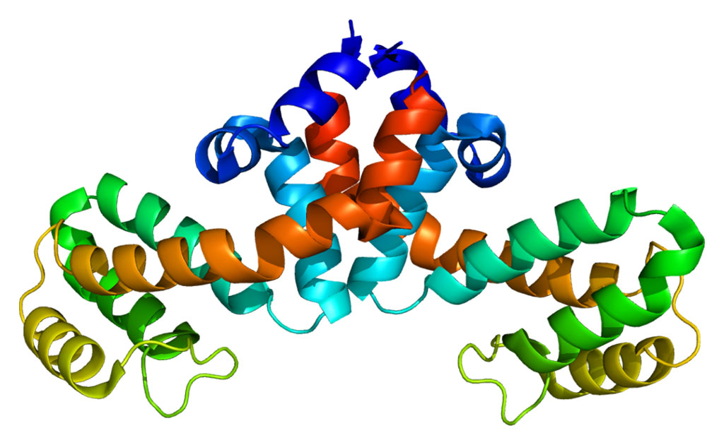
Duchenne muscular dystrophy (DMD) is caused by mutations in the gene that encodes dystrophin, a protein crucial for maintaining muscle cell integrity and function, and the subsequent disruption of the dystrophin-associated protein complex (DAPC). The mutation occurs on the X-chromosome, and the disease effects about one of every 3,500 boys whose muscle function is so degraded that they die usually before reaching the age of 30. The majority of DMD mutations are deletions that prematurely terminate the dystrophin protein. Deletions of exon 50 of the dystrophin gene are among the most common single exon deletions causing DMD. Such mutations can be corrected by skipping exon 51, thereby restoring the dystrophin reading frame.

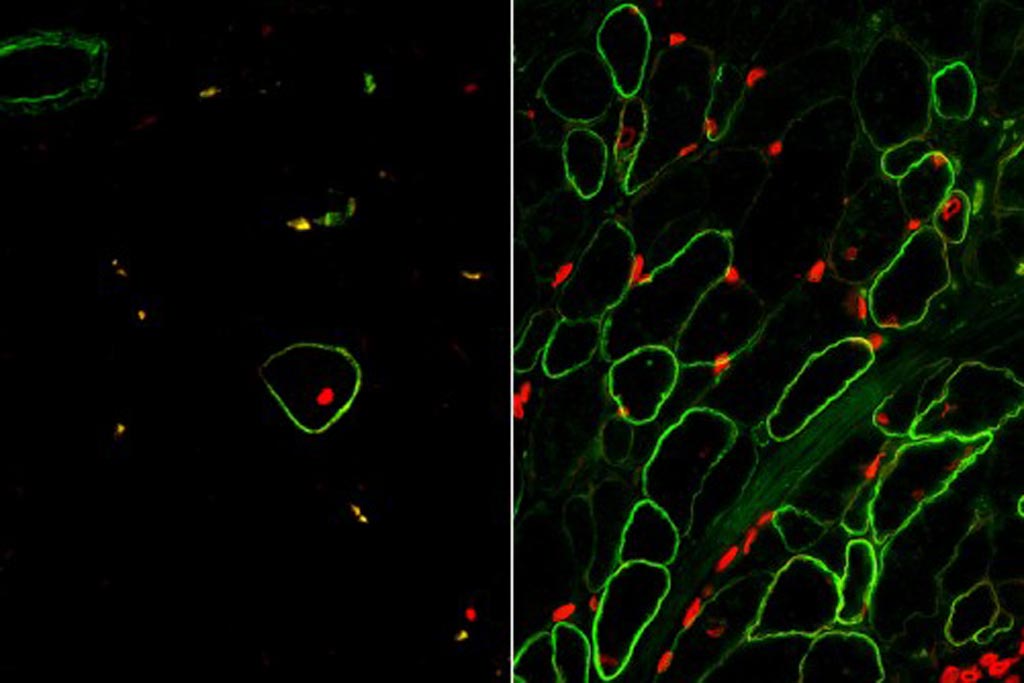
Image: Skeletal muscle cells isolated using the ERBB3 and NGFR surface markers (right) restore human dystrophin (green) after transplantation significantly greater than previous methods (left) (Photo courtesy of the University of California, Los Angeles and Nature Cell Biology).
Human pluripotent stem cells (hPSCs) can be directed to differentiate into skeletal muscle progenitor cells (SMPCs). However, the myogenicity of hPSC-SMPCs relative to human fetal or adult satellite cells has been unclear, and it has been observed that hPSC-SMPCs derived by directed differentiation were less functional in vitro and in vivo compared to human satellite cells.
To improve the efficiency of stem cell-derived muscle tissue, investigators at the University of California, Los Angeles (USA) first used RNA sequencing to identify cell surface receptors that marked fetal-derived muscle cell populations. They chose the proteins ERBB3 (receptor tyrosine-protein kinase erbB-3) and NGFR (low-affinity nerve growth factor receptor), and used them as markers in subsequent cell selection procedures.
The investigators found that stem cell-derived muscle cell populations were immature, but that inhibition of transforming growth factor-beta (TGF-beta) signaling during differentiation improved fusion efficiency, ultrastructural organization, and the expression of adult myosins. In the next step, cells from DMD patients were induced into becoming pluripotent stem cells. The investigators then corrected the DMD-causing genetic mutation using the gene editing technology CRISPR/Cas9.
CRISPR/Cas9 is regarded as the cutting edge of molecular biology technology. CRISPRs (clustered regularly interspaced short palindromic repeats) are segments of prokaryotic DNA containing short repetitions of base sequences. Each repetition is followed by short segments of "spacer DNA" from previous exposures to a bacterial virus or plasmid. Since 2013, the CRISPR/Cas9 system has been used in research for gene editing (adding, disrupting, or changing the sequence of specific genes) and gene regulation. By delivering the Cas9 enzyme and appropriate guide RNAs (sgRNAs) into a cell, the organism's genome can be cut at any desired location. The conventional CRISPR/Cas9 system is composed of two parts: the Cas9 enzyme, which cleaves the DNA molecule and specific RNA guides that shepherd the Cas9 protein to the target gene on a DNA strand. Efficient genome editing with Cas9-sgRNA in vivo has required the use of viral delivery systems, which have limitations for clinical applications.
The investigators used the ERBB3 and NGFR surface markers to isolate CRISPR-modified, stem cell-derived skeletal muscle cells, which were then injected into mice at the same time as a TGF-beta inhibitor. Results published in the December 18, 2017, online edition of the journal Nature Cell Biology revealed that this enrichment and maturation strategy restored dystrophin in hundreds of dystrophin-deficient myofibers after engraftment of CRISPR/Cas9-corrected DMD hPSC-SMPCs.
“We have found that just because a skeletal muscle cell produced in the lab expresses muscle markers, does not mean it is fully functional,” said senior author Dr. April Pyle, associate professor of microbiology, immunology, and molecular genetics at the University of California, Los Angeles. “For a stem cell therapy for Duchenne to move forward, we must have a better understanding of the cells we are generating from human pluripotent stem cells compared to the muscle stem cells found naturally in the human body and during the development process. The results were exactly what we had hoped for. This is the first study to demonstrate that functional muscle cells can be created in a laboratory and restore dystrophin in animal models of Duchenne using the human development process as a guide.”
Related Links:
University of California, Los Angeles


