Novel Culture Method for Activation of Cancer-Fighting T-Cells
By LabMedica International staff writers
Posted on 02 Mar 2017
A novel in vitro culture method enables disease fighting immune T-cells to overcome cancer's immunosuppressive effect in order to recognize and attack tumor cells upon being returned to the body.Posted on 02 Mar 2017
Development of effective adoptive immunotherapy for many types of human cancer has been slow, often due to difficulties achieving robust expansion of natural tumor-specific T-cells from peripheral blood. Investigators at the Mayo Clinic and the University of Washington hypothesized that antigen-driven T-cell expansion might best be triggered in vitro by acute activation of innate immunity to mimic a life-threatening infection.

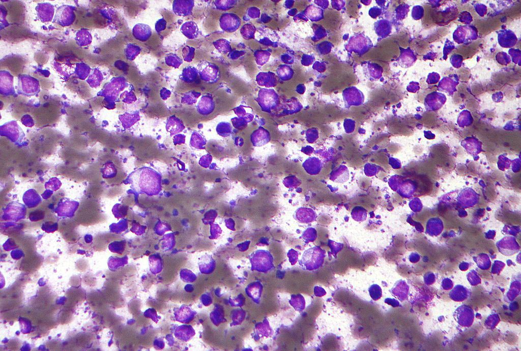
Image: A novel in vitro culture method enables disease fighting immune T-cells to overcome cancer\'s immunosuppressive effect (Photo courtesy of the Mayo Clinic).
To examine this theory, they subjected unfractionated peripheral blood mononuclear cells (PBMC) to a two-step culture regimen, first synchronizing their exposure to exogenous antigens with aggressive surrogate activation of innate immunity, followed by gamma-chain cytokine-modulated T-cell hyperexpansion.
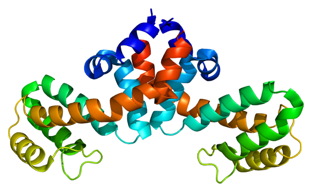
In the first step, the PBMC culture was treated with granulocyte-macrophage colony-stimulating factor (GM-CSF) plus paired Toll-like receptor agonists (resiquimod and LPS), which stimulated abundant IL-12 and IL-23 secretion. At this point the culture was exposed to various tumor antigens including MUC1 (Mucin 1, cell surface associated), a protein expressed by a large majority of cancers, including breast, pancreatic, lung, colorectal, ovarian, kidney, bladder, and multiple myeloma. Also included were HER2/neu (human epidermal growth factor receptor 2), a protein present in one-quarter to half of many types of cancer, and CMVpp65, a protein present in half of primary brain tumors.
In the second step, exposure to exogenous IL-7 or IL-7+IL-2 produced selective and sustained expansion of both CD4+ and CD8+ peptide-specific T-cells with a predominant interferon-gamma-producing T1-type, as well as the antigen-specific ability to lyse tumor targets. The investigators reported in the February 14, 2017, issue of the journal Oncotarget that it only took about three weeks to grow out cultures of natural T- cells able to recognize and target cancers expressing these proteins.
“Even though it is relatively easy to collect billions of T-cells directly from patient blood, it has historically proved difficult or impossible to unleash those T-cells’ natural ability to recognize and target cancer cells,” said senior author Dr. Peter Cohen, an immunotherapist at the Mayo Clinic. “We are pleased to help other investigators implement our culture method for their own cancer-associated proteins of interest.”


