Synthetic Printed Implants Prompt Spinal Regeneration in Model
By LabMedica International staff writers
Posted on 31 Jan 2019
The potential use of three-dimensional (3D) printing to produce replacement components for the repair of spinal damage was demonstrated in a rat model system.Posted on 31 Jan 2019
Up to now, three-dimensional printing of central nervous system (CNS) structures has not been accomplished, possibly owing to the complexity of CNS architecture. To rectify this situation, investigators at the University of California, San Diego (USA) used a three-dimensional microscale continuous projection printing method (MuCPP) to create a complex CNS structure for regenerative medicine applications in the spinal cord.

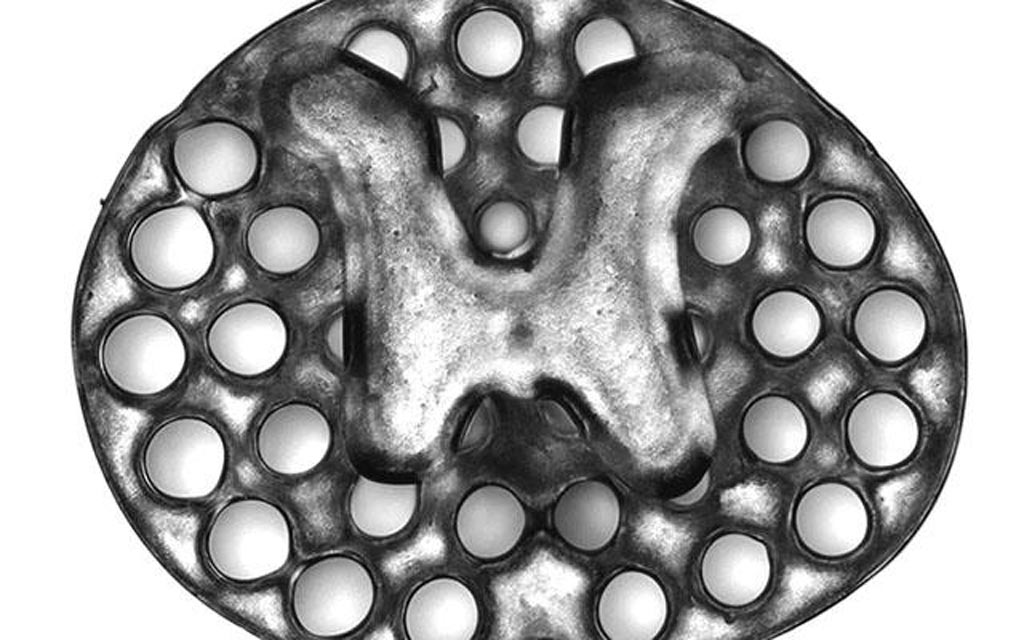
Image: A three-dimensional printed, two-millimeter implant used as scaffolding to repair spinal cord injuries in rats. The circles surrounding the H-shaped core are hollow portals through which implanted neural stem cells can extend axons into host tissues (Photo courtesy of Jacob Koffler and Wei Zhu, University of California, San Diego).
The MuCPP method enabled printing of three-dimensional biomimetic hydrogel scaffolds that were tailored to the dimensions of the rodent spinal cord. This process required only 1.6 seconds and was scalable to human spinal cord sizes and lesion geometries. In this regard, four-centimeter-sized implants modeled from MRI scans of actual human spinal cord injuries were printed within 10 minutes. The printed scaffolds contained dozens of 200-micrometer-wide channels that guided neural stem cell and axon growth along the length of the spinal cord injury.
The investigators tested the ability of MuCPP three-dimensional-printed scaffolds loaded with neural progenitor cells (NPCs) to support axon regeneration and form new "neural relays" across sites of complete spinal cord injury in vivo in rodents.
They reported in the January 14, 2019, online edition of the journal Nature Medicine that injured host axons regenerated into three-dimensional biomimetic scaffolds and synapsed onto NPCs implanted into the device. Implanted NPCs in turn extended axons out of the scaffold and into the host spinal cord below the injury to restore synaptic transmission and significantly improve the animal's ability to move.
"In recent years and papers, we have progressively moved closer to the goal of abundant, long-distance regeneration of injured axons in spinal cord injury, which is fundamental to any true restoration of physical function," said senior author Dr. Mark Tuszynski, professor of neuroscience at the University of California, San Diego.
Related Links:
University of California, San Diego


